Faire une echographie à Paris
Ou faire une échographie à Paris ?

What is an ultrasound scan?
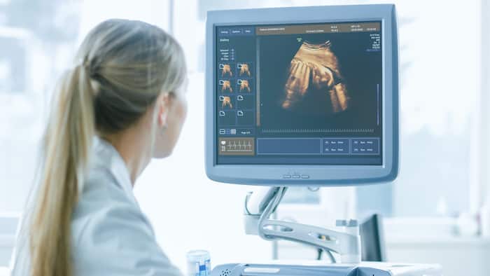
L’échographie est un examen sans douleur et sans radiation qui exploite les propriétés des ultrasons, n’ayant aucun impact sur la santé du patient. La technologie est similaire à celle utilisée par le sonar et le radar, qui aident les militaires à détecter les avions et les navires. Une échographie offre à votre médecin la possibilité d’examiner les problèmes des organes, des vaisseaux et des tissus sans nécessiter d’incision.
Faire une échographie est une technique d’imagerie médicale non invasive utilisée pour établir un diagnostic précis. Elle permet de visualiser en temps réel les organes et tissus, comme dans l’échographie mammaire, un type d’examen essentiel pour détecter des anomalies. Dans un centre d’échographie ou un centre de radiologie, des médecins généralistes ou des radiologues spécialisés effectuent ces examens pour assurer une prise en charge optimale. La sécurité sociale couvre en grande partie ces examens de radiologie, garantissant ainsi un accès facilité à ces soins essentiels pour les patients.
Contrairement à d’autres techniques d’imagerie, l’échographie n’utilise aucun rayonnement. Pour cette raison, c’est la méthode préférée pour visualiser un fœtus en développement pendant la grossesse.
Où faire une échographie à Paris ?
Pour réaliser une échographie à Paris, plusieurs options s’offrent à vous. Vous pouvez consulter des centres d’imagerie médicale spécialisés, des hôpitaux ou encore des cliniques privées. Il est essentiel de choisir un établissement équipé d’appareils modernes et d’un radiologue expérimenté pour assurer la qualité et la précision des résultats. Notre centre d’imagerie médicale IMPC proposent aux patients:
1- des appareils très avancés,
2- ainsi qu’une équipe pluridisciplinaire assurant une prise en charge complète et personnalisée en combinant l’expertise de spécialistes variés pour des diagnostics précis et adaptés. Cette approche optimise la qualité des soins et le suivi des patients
How does an ultrasound scan work?
You'll be lying down in a darkened room to make it easier to read the images on the video screen, which you can follow on a second screen dedicated to the patient.
A gel will be applied to the skin to enable ultrasound transmission.
The examination provides dynamic moving images, controlled on a screen.
What is it all about?
Ultrasound uses ultrasound emitted by a probe and transmitted through the tissues, which reflect them, to form an image of the region examined. It can be coupled with a kind of radar to study vessels (Doppler).
Is it necessary to inject a product?
NO, the test is performed without injection (prick) and is painless.
Your results
Un premier commentaire pourra vous être donné juste après l’examen, il ne s’agira là que d’une première approche, car les images doivent ensuite faire l’objet d’un compte rendu écrit qui sera disponible dans les meilleurs délais. Il est normal que vous vous posiez des questions sur l’examen que vous êtes amené à passer. Nous espérons y avoir répondu. N’hésitez pas à nous interroger à nouveau pour tout renseignement complémentaire.
Pelvic ultrasound
During the pelvic ultrasound, do not urinate for 3 hours before the examination, or if you have urinated, drink 1 liter of water 1 hour beforehand. To be in immediate contact with the area under examination and improve image quality, you may be offered the option of placing a probe covered with sterile protection in the rectum or vagina. Very rarely, the introduction of the probe may cause transient, non-serious discomfort. Not all anatomical structures can be visualized by ultrasound. The liver, spleen, kidneys, bladder, uterus and ovaries are particularly well studied. The study of the kidneys and bladder as well as gynecological organs often requires a full bladder examination. Muscles and tendons are also well studied, but osteoarticular ultrasound requires no special preparation of the patient.
Qu’est-ce qu’une échographie abdomino-pelvienne ?
An abdomino-pelvic ultrasound enables visualization of the abdominal and pelvic organs. This examination is performed through the abdominal wall or by endocavity.
Pourquoi faire une échographie abdomino-pelvienne ?
- Diagnosing a disease : tumor detection (e.g. ovarian cyst), infection (salpingitis, cholecystitis, pyelonephritis for example), a calculation (vesicular lithiasiskidney, for example)... ;
- Keeping pace of a pathology (cancer surveillance, treatment for a endometriosis…) ;
- Tracking growth and study the proper development of fetal organs during the pregnancy ;
- Guiding a surgeon through certain operationsetc.
Quand faire une échographie abdomino-pelvienne ?
Pelvic pain, i.e. pain felt in the lower abdomen, which generally involves the uterus, bladder and rectum, and abdominal pain, which can take the form of cramping, burning or throbbing, can in some cases reveal abnormalities that require follow-up, treatment or intervention.
Any pelvic or abdominal pain persisting for more than 6 weeks should be reported to a radiologist.
Qu’est-ce qu’une échographie abdominale ?
It's the ideal examination for exploring solid organs: the liver, gallbladder and bile ducts, kidneys and spleen.
Why do I need an abdominal ultrasound?
Abdominal ultrasound provides a wealth of information, much richer than standard radiography on the abdominal organs.
The main organs explored by abdominal ultrasound and the abnormalities are :
Gallbladder and bile ducts: stones, dilatation, tumors, abscesses, inflammation of the wall ;
Liver: size and homogeneity (cirrhosis, steatosis), cysts, abscesses, tumors, metastases of non-hepatic cancers;
Spleen: size, swelling, suspected rupture;
Pancreas: cyst, pancreatitis due to gallstones, cancer;
Kidneys: stones, dilated pelvis or ureters, tumours, atrophy, malposition, cysts, malformations;
Aorta and inferior vena cava;
The muscular wall of the abdomen.
Ultrasound may also show abnormal lymph nodes, ascites, blood or suspicious masses in the abdominal cavity.
Quand faire une échographie abdominale ?
If abdominal pain persists beyond 6 weeks, an ultrasound scan may be prescribed.
Do you have to fast before theabdominal ultrasound ?
To optimize the examination, you must be fasting and not have had a drink or smoked for at least 3 hours.
Qu’est-ce qu’une échographie obstétricale ?
An obstetrical ultrasound is a medical radiological examination that visualizes images of the baby (embryo or fetus) in a pregnant woman, as well as the mother's uterus and ovaries. It uses no ionizing radiation, has no known harmful effects and is the preferred method for monitoring pregnant women and their unborn babies.
That said, the use of obstetrical ultrasound is unquestionably one of the most essential examinations during pregnancy.
Pourquoi faire une échographie obstétricale ?
Aobstetrical ultrasound is a useful clinical examination for :
- Establishing the presence of a living embryo/foetus
- Estimating pregnancy age
- Diagnosing congenital anomalies of the fetus
- Assessing fetal position
- Assessing the position of the placenta
- Determining multiple pregnancies
- Determining the amount of amniotic fluid around the baby
- Check for cervical opening or shortening
- Assessing fetal growth
- Assessing fetal well-being
What's more, its usefulness is enhanced by the fact that this ultrasound assiduously accompanies the woman's entire pregnancy, paying particular attention to the baby's development right up to birth.
Quand faire une échographie obstétricale ?
In fact, three obstetrical ultrasounds are required during pregnancy.
- A dating ultrasound: this takes place at the very beginning. This is used to mark the date and make an appointment for ultrasound to be performed between 11 weeks and 1 day and 13 weeks and 6 days of amenorrhea.
- La 2e Ultrasound is a much more in-depth study of fetal morphology and adnexa (Doppler, placenta).
- La 3e The ultrasound will determine the baby's development, fetal weight, fetal position and placental position. The heart and brain will be more visible than on the 2e quarter.
RDV ECHOGRAPHIE en ligne
Make an appointment for an ultrasound scan at one of our centers:
Radiology Montmartre 75002
Pôle Santé Bergère 75009
Blomet radiology 75015
Radiology Belleville 75020
