IRM mammaire: prévenir le cancer du sein

RDV IRM MAMMAIRE en ligne sur Doctolib
Prenez rendez-vous pour un IRM dans nos centres
Prise de rendez-vous IRM – Paris 75002
Prise de rendez-vous IRM – Paris 75009 – Pôle Santé Bergère
Prise de rendez-vous IRM – Paris 75009 – Clinique Drouot
Prise de rendez-vous IRM – Paris 75015

Comment se passe une IRM mammaire dans notre centre ?
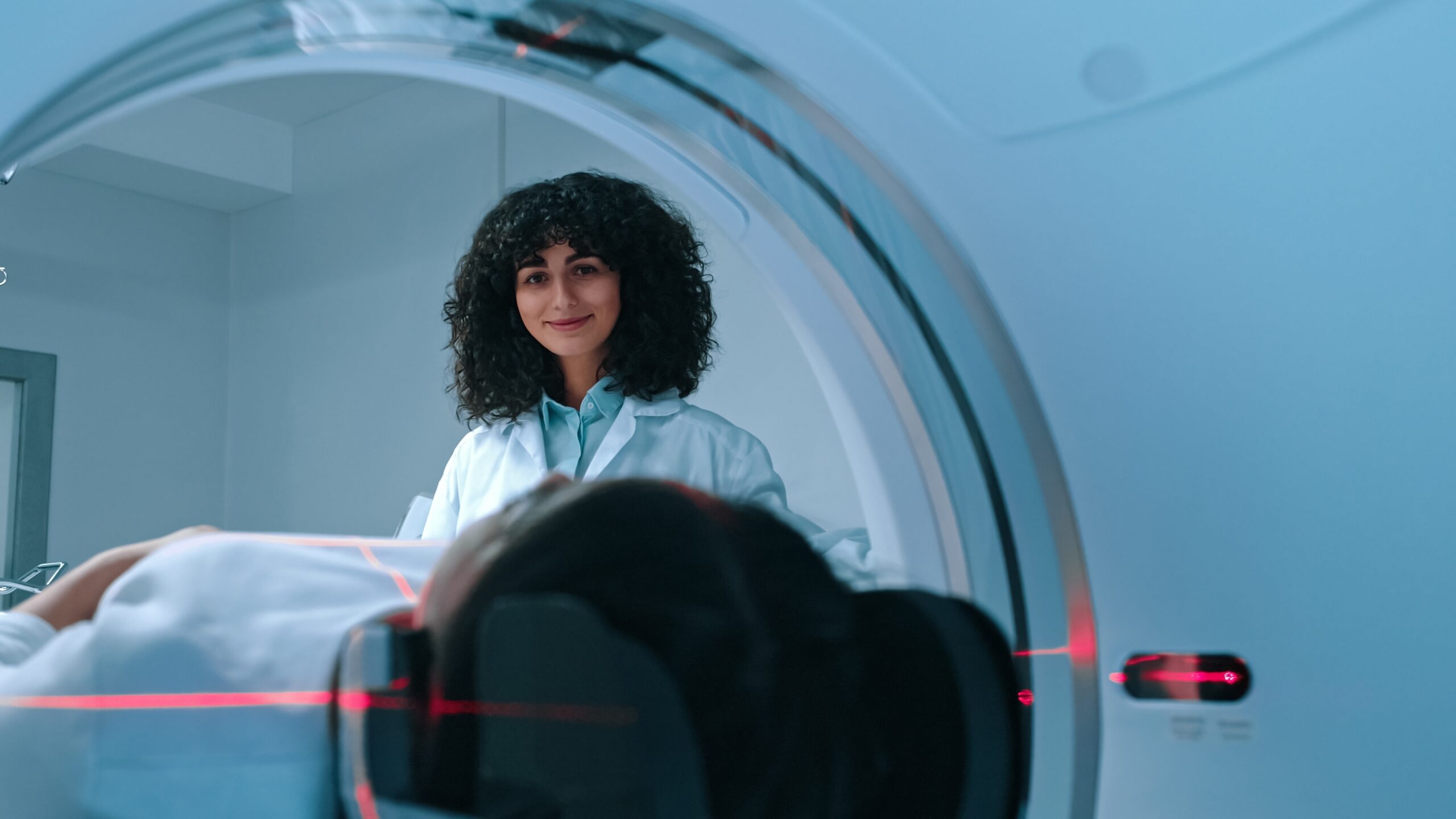
L’IRM mammaire se déroule dans un tunnel d’IRM. La patiente est installée allongée sur le ventre, les seins positionnés dans des cavités adaptées à une antenne spécifique.
La particularité de nos centres IMPC est de disposer d’appareils semi-ouverts, qui facilitent l’examen pour les patientes claustrophobes et améliorent leur confort. Tout au long de la procédure, le manipulateur-radio et le médecin radiologue sont présents pour guider et rassurer la patiente.
Quelles sont les étapes de l’examen ?
Accueil et préparation : la patiente est appelée en salle d’attente et installée dans une cabine. Elle doit retirer tout objet métallique (bijoux, montre, piercing).
Vêtements : il est demandé d’enlever le soutien-gorge et de conserver un tee-shirt ou une blouse médicale.
Installation : la patiente est conduite dans la salle d’IRM, puis allongée sur la table en position ventrale. Les seins sont placés dans une antenne mammaire conçue pour recueillir le signal.
Injection : une injection de produit de contraste peut être réalisée par voie intraveineuse pour améliorer la visualisation des tissus et vaisseaux.
Acquisition des images : la table glisse doucement dans le tunnel, l’examen dure environ 25 à 30 minutes. La patiente doit rester immobile, tout en étant équipée d’un casque ou de bouchons d’oreilles pour atténuer le bruit.
Results : après l’examen, la patiente retourne en salle d’attente. Le médecin radiologue analyse les images et peut donner une première interprétation sous 20 minutes.
La patiente repart avec ses clichés (papier et CD), et le compte rendu est remis en main propre ou téléchargeable sur notre site sécurisé.
Particularité IMPC
Nos centres d’imagerie médicale sont équipés d’IRM semi-ouvertes, qui limitent la sensation de confinement et permettent aux patientes anxieuses ou claustrophobes de passer l’examen dans de meilleures conditions.
L’IRM mammaire est-elle efficace ?
Oui. L’IRM mammaire est aujourd’hui reconnue comme l’examen le plus sensible pour le diagnostic du cancer du sein.
-
Sa sensibilité dépasse 90 % pour les cancers infiltrants, contre environ 70 % pour la mammographie, surtout chez les femmes aux seins denses (Radiology, 2021).
-
Elle permet de détecter des lésions multifocales ou bilatérales souvent manquées par les autres examens.
-
Elle visualise la néo-angiogenèse tumorale (développement anormal de vaisseaux sanguins), un critère essentiel dans l’évaluation de l’agressivité des cancers.
Selon le SNITEM (2023), près de 300 000 IRM mammaires sont réalisées chaque année en France, soulignant l’importance croissante de cet examen dans la prise en charge des patientes.
Ce que l’IRM mammaire permet de détecter
L’IRM mammaire offre une exploration complète et précise des tissus mammaires.
Les tumeurs et cancers
Elle repère :
-
from lésions malignes très précoces, parfois invisibles à la mammographie,
-
from cancers multifocaux (plusieurs foyers dans un même sein),
-
from cancers bilatéraux, touchant les deux seins simultanément,
-
from anomalies de rehaussement après injection de contraste, typiques d’une néo-angiogenèse tumorale.
Les masses bénignes
Elle distingue clairement :
-
from kystes simples ou complexes,
-
from adénofibromes, souvent difficiles à caractériser en mammographie.
La surveillance après traitement
Elle permet de :
-
rechercher une récidive locale après chirurgie ou radiothérapie,
-
contrôler la réponse à la chimiothérapie néoadjuvante, en mesurant la réduction de la tumeur.
Les prothèses et implants mammaires
L’IRM est la méthode de référence pour :
-
détecter les ruptures intracapsulaires (invisibles à l’échographie),
-
identifier les plis ou anomalies de forme,
-
analyser les réactions inflammatoires péri-prothétiques.
Étendue des explorations mammaires par IRM
L’IRM mammaire offre une analyse précise de la glande mammaire, des canaux galactophores, du tissu conjonctif et des ganglions axillaires.
Elle permet de détecter des tumeurs précoces, de contrôler l’intégrité des implants et d’évaluer les anomalies dans les seins denses.
Elle peut également guider une biopsie ou orienter une chirurgie conservatrice, en complément de la mammographie et de l’échographie.
Qu’est-ce qu’une IRM mammaire ?
ABreast MRI, également appelée Breast MRI, est un examen d’imagerie médicale avancé permettant d’explorer avec une très grande précision la structure interne des seins.
Contrairement à la mammographie, elle n’utilise pas de rayons X mais repose sur l’association d’un champ magnétique puissant et d’ondes radio.
Cet examen est réalisé en deuxième intention : il n’est pas destiné au dépistage systématique, mais intervient lorsque la mammographie ou l’échographie n’ont pas permis de fournir un diagnostic complet.
Grâce à l’IRM, le médecin radiologue peut obtenir des images en coupes successives des tissus mammaires, évaluer la vascularisation des lésions et détecter des anomalies parfois invisibles avec les autres techniques.
Pourquoi faire une IRM mammaire ?
L’IRM mammaire est un outil indispensable dans de nombreuses situations cliniques. Elle est notamment indiquée pour :
-
Surveiller les femmes à haut risque génétique (mutations BRCA1, BRCA2, antécédents familiaux de cancer du sein).
-
Évaluer un cancer du sein récemment diagnostiqué afin de déterminer son extension exacte.
-
Rechercher un cancer non détecté à la mammographie ou à l’échographie, surtout chez les femmes aux seins denses.
-
Contrôler l’intégrité des implants ou prothèses mammaires, notamment en cas de suspicion de rupture.
-
Préparer une chirurgie conservatrice, en localisant avec précision la ou les lésions.
-
Évaluer la réponse à une chimiothérapie néoadjuvante, en vérifiant la régression tumorale avant chirurgie.
Ainsi, l’IRM mammaire ne remplace pas les examens standards mais les complète pour offrir une vision globale et sécuriser le parcours de soins.
Préparation à l’examen
Pour garantir la fiabilité des images :
-
L’examen est idéalement programmé entre le 7e et le 14e jour du cycle menstruel, afin de limiter l’influence hormonale.
-
The world of medical imaging has made significant strides in diagnosing and understanding the human body. As one of these technologies, Magnetic Resonance Imaging (MRI) offers detailed images of internal structures without requiring invasive procedures. In this article, we discuss the crucial role of lumbar spine MRI in diagnosing conditions affecting the lower back. prise de sang peut être prescrite pour vérifier la fonction rénale avant l’injection de produit de contraste.
-
La patiente doit signaler tout implant métallique, pacemaker ou antécédent allergique.
-
En cas de claustrophobie, une adaptation de la prise en charge est possible.
Injection de produit de contraste
La majorité des IRM mammaires nécessitent une injection intraveineuse de gadolinium. Ce produit :
-
permet de mieux visualiser les vaisseaux et anomalies,
-
améliore la détection des lésions infiltrantes,
-
aide à différencier les masses bénignes et malignes.
Les réactions allergiques sont extrêmement rares (moins de 0,1 % selon la HAS, 2021).
Sécurité et contre-indications
L’IRM mammaire est un examen sûr et non invasif :
-
aucun rayonnement ionisant,
-
examen compatible avec la grossesse sans injection (après accord médical),
-
effets secondaires très rares.
Contre-indications absolues :
-
pacemaker ou défibrillateur non compatible,
-
neurostimulateur, implant auditif ou métallique non certifié IRM.
En cas d’insuffisance rénale sévère, l’injection de gadolinium peut être évitée.
Tableau comparatif : IRM mammaire vs mammographie
| Critère | Breast MRI | Mammography |
|---|---|---|
| Sensibilité diagnostique | > 90 % (cancers infiltrants) | ≈ 70 % (moins efficace si seins denses) |
| Rayons X | Aucun | Oui |
| Visualisation bilatérale | Oui, simultanée | Oui, mais moins précise |
| Duration | 25–30 minutes | 10–15 minutes |
| Indications principales | Haut risque, bilan d’extension, prothèses | Dépistage standard, suivi courant |
Pourquoi choisir IMPC pour votre IRM mammaire ?
En choisissant IMPC, vous bénéficiez :
-
d’un réseau de 10 centres d’imagerie médicale à Paris,
-
d’appareils IRM de dernière génération,
-
from radiologues spécialisés en imagerie mammaire,
-
d’une prise de rendez-vous rapide et simple via Doctolib,
-
d’un accès en ligne sécurisé à vos résultats.
Notre priorité : offrir un examen fiable, dans un cadre rassurant et avec un suivi personnalisé.
FAQ : Les questions les plus courantes sur l’IRM mammaire
Combien de temps dure une IRM mammaire ?
En moyenne 25 à 30 minutes, parfois un peu plus si un produit de contraste est injecté.
Faut-il être à jeun pour une IRM mammaire ?
Non, sauf si le radiologue le précise, notamment en cas d’injection de contraste.
L’IRM mammaire est-elle douloureuse ?
Non, l’examen est indolore. Seule l’injection peut occasionner une légère gêne.
Peut-on faire une IRM mammaire enceinte ?
Oui, mais sans injection, et uniquement si le bénéfice médical est jugé supérieur au risque.
Quand reçoit-on les résultats ?
Le compte rendu est disponible rapidement, avec clichés papier, CD et accès en ligne sécurisé.
L’IRM mammaire peut-elle remplacer la mammographie ?
Non. Elle est complémentaire et prescrite en cas de doute ou de risque élevé.
Nos spécialistes :
-
Nabil Jomaah
Sources scientifiques
-
Haute Autorité de Santé (HAS) – Sécurité des examens IRM et produits de contraste (2021)
-
Inserm – Cancer du sein : dépistage et facteurs de risque (2022)
-
Société Française de Radiologie – Recommandations en imagerie mammaire
-
Radiology (2021) – MRI sensitivity in invasive breast cancer
Dernière mise à jour : 30 août 2025
Dr Corinne Bordonné
