How does a mammogram work?
Mammography in Paris
What is mammography?
A mammogram is a low-dose X-ray of breast tissue. Health professionals use mammograms to look for the first signs of breast cancer before symptoms appear. This is called a screening mammogram. They also use mammography to look for abnormalities if you develop a new symptom, such as a lump, pain, nipple discharge or changes in the skin of the breast. This is called a diagnostic mammogram.
Apart from skin cancer, breast cancer is the most common cancer affecting people designated as female at birth, accounting for 49,000 new cases each year in France. Although breast cancer treatment therapies continue to improve and have contributed to a reduction in cancer-related deaths, early diagnosis through screening mammograms has a greater overall impact on survival rates.
Most mammography results are benign, or non-cancerous. In fact, less than 1 in 10 people requiring further testing after a mammogram has cancer.

Why have a mammogram?
Most breast cancers are neither painful nor palpable, as they are located deep within the breast and are small in size. Mammography can detect a small cancer at an early stage of development, thereby increasing the patient's chances of being cured, as the size of the lesion is a major prognostic factor. In this case, it's a "screening" mammogram.
Sometimes mammography is performed because the patient or the doctor has palpated an abnormal area in a breast. The mammographic appearance will help to determine whether the patient should be reassured or whether further investigations should include a breast biopsy.
How does a mammogram work?
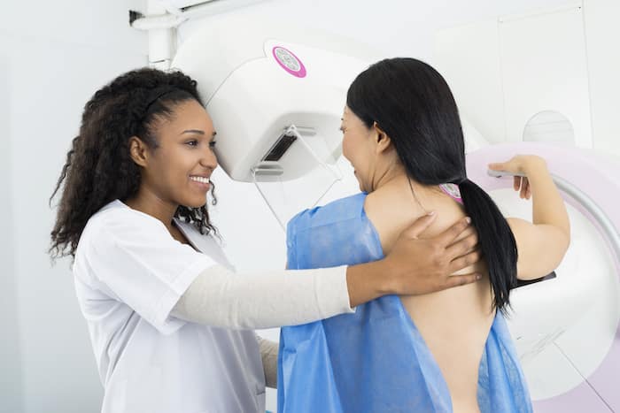
The patient is bare-chested. The operator gently positions the patient against the mammograph. The breast is gradually compressed so that it does not move when the image is taken. Each breast is compressed twice to obtain a "front" and "oblique" view of each side. Each compression lasts a few seconds.
Do breast implants interfere with mammograms?
Having silicone or saline breast implants and the resulting scar tissue make it more difficult for radiologists to see all your breast tissue and possible problems on regular mammograms.
To help the radiologist see as much breast tissue as possible, people with implants usually have two additional images taken of each breast in addition to the standard four images taken during a screening mammogram. These additional images are called implant displacement views (IDVs).
For VDI, the technologist gently pushes your breast implant against your chest wall, pulls your breast forward over it and then compresses your breast. This allows better imaging of the front of each breast.
It is important to inform the mammography center that you have breast implants when you book your mammogram, and to inform your technologist on the day of your mammogram.
Why have a breast ultrasound after a mammogram?
The radiologist is free to decide whether or not to perform a breast ultrasound in addition to mammography.
It will be carried out in two circumstances:
- most frequently because the breast is considered "dense" on mammography, which hinders the detection of abnormalities. In this case, an additional ultrasound scan improves detection sensitivity.
- if an abnormality is detected in mammography and the radiologist wishes to explore it further in ultrasound.
When the breasts are not very dense, and the radiologist has previous images for comparison, breast ultrasound is generally not necessary.
What is a breast ultrasound?
Ultrasound breastThis non-irradiating procedure is performed as a first-line treatment for young women (under 30), and as a complement to mammography in the case of dense or heterogeneous breasts, or if there is an abnormality to be characterized.
What is a mammary cyst?
A breast cyst is a "pocket of water". It's a very common breast lesion, and completely benign (harmless) if considered "simple". In this case, no special surveillance is recommended, as the cyst is not dangerous.
If the cyst is painful or unsightly due to its size, the radiologist can perform a "puncture-evacuation" under ultrasound control, i.e. he gently evacuates the liquid contained in the cyst using a small syringe and a fine needle, which he can guide precisely under ultrasound control.
If the cyst is deemed "atypical" by the radiologist, he or she may decide to monitor it with an ultrasound scan in 4 months' time, or to remove the cyst and send the fluid to the laboratory to determine whether or not it is dangerous.
What is a breast nodule?
A "nodule" is a small "solid" mass in the breast, i.e. it doesn't contain water like a cyst, but tissue. This tissue may be totally benign, made up of fibers and glands (fibroadenoma), or malignant (therefore dangerous). The radiologist judges the degree of suspicion on the basis of precise mammo-echographic semiological criteria, and decides whether to puncture or monitor the nodule.
Why are my breasts compressed during a mammogram?
Compression is necessary to eliminate overlapping tissue and obtain a clear image of the inside of the breast.
Is mammography painful?
While some women find the examination uncomfortable, the pain is generally short-lived.
What are the different types of mammograms?
Il existe deux principaux types de mammographies :
- 2D digital mammography.
- 3D digital mammography (digital breast tomosynthesis).
Digital mammography
Today, digital mammography has replaced conventional (film) mammography, also known as analog mammography. Both digital and conventional mammography use X-rays to produce an image of your breast. The difference is that the image is stored directly on film in conventional mammography, while digital mammography provides an electronic image stored as a computer file. Digital mammography enables healthcare professionals to save the file electronically, and to evaluate and share images more easily.
A digital mammogram usually involves at least two images of each breast taken from different angles, usually top-to-bottom and side-to-side, and offers a two-dimensional (2D) view.
3D mammography
3D mammography, also known as digital breast tomosynthesis (DBT), is a newer type of mammography in which each breast is compressed once and a machine takes several low-dose X-rays as it moves in an arc over your breast. A computer then stitches the images together, allowing medical professionals to see your breast tissue more clearly in three dimensions.
Numerous studies have shown that 3D mammography increases the detection of cancers, including low-grade cancers, and reduces false-positive rates. Given these advantages, 3D mammography, for both screening and diagnosis, is fast becoming the preferred option for mammography.
What's the difference between screening mammography and diagnostic mammography?
A screening mammogram is a routine (usually annual) mammogram that healthcare professionals recommend to look for signs of cancer or abnormal breast tissue before you have any symptoms. Screening mammography helps in the early detection of breast cancer. Early detection enables early treatment, which can be more effective than if the cancer is discovered at a later stage.
A routine screening mammogram usually includes at least two images of each breast taken from different angles, typically top to bottom and side to side. If you have breast implants, additional images will be required.
Health professionals prescribe a diagnostic mammogram if a screening mammogram shows abnormal tissue or if there is a new breast problem.
Although both types of mammography use the same machines, diagnostic mammography employs additional imaging techniques, such as localized compression, additional angles or enlarged views, and is supervised by the radiologist at the time of the study.
How does a mammogram work?
A mammogram uses an X-ray machine designed specifically to examine breast tissue. The machine produces X-rays at lower doses than those used to examine your bones.
During a mammogram, you place your breast on a support plate attached to the X-ray machine. A technologist then compresses your breast with a parallel plate called a paddle. The machine produces X-rays that pass through your breast to a detector on the opposite side. The detector transmits electronic signals to a computer to form a digital image. These images are called mammograms.
Breast compression is necessary during mammography to keep your breast still and minimize movement, which can blur X-rays. Compression also evens out the shape of your breast so that X-rays can travel a shorter path to reach the detector. This results in a lower radiation dose and improved image quality.
What's the difference between screening mammography and diagnostic mammography?
A screening mammogram is a routine (usually annual) mammogram that healthcare professionals recommend to look for signs of cancer or abnormal breast tissue before you have any symptoms. Screening mammography helps in the early detection of breast cancer. Early detection enables early treatment, which can be more effective than if the cancer is discovered at a later stage.
A routine screening mammogram usually includes at least two images of each breast taken from different angles, typically top to bottom and side to side. If you have breast implants, additional images will be required.
Health professionals prescribe a diagnostic mammogram if a screening mammogram shows abnormal tissue or if there is a new breast problem.
Although both types of mammography use the same machines, diagnostic mammography employs additional imaging techniques, such as localized compression, additional angles or enlarged views, and is supervised by the radiologist at the time of the study.
At what age should I have a mammogram?
This is a subject of debate among experts. In general, women are encouraged to start screening mammograms between the ages of 40 and 50. However, if you have a family history of breast cancer, it's advisable to consult your doctor to determine the right age to start.
Breast cancer screening
Mammography is one of the most effective ways of detecting breast cancer at an early stage, well before symptoms appear. By identifying tumors at an early stage, the chances of successful treatment increase considerably.
RDV MAMMOGRAPHIE en ligne
Make an appointment for a mammogram at one of our centers:
Radiology Montmartre 75002
Pôle Santé Bergère 75009
Blomet radiology 75015
Radiology Belleville 75020
How do I prepare for a mammogram?
It is not necessary to fast or stop taking any medication before a mammogram. It is preferable not to put any cream on the skin or deodorant under the armpit. Piercings should be removed.
The patient is asked to bring :
- prescription (sometimes important clinical information is specified by the doctor)
- previous mammograms
At what age should I have a mammogram?
In France, mammograms are performed every 2 years as part of the organized breast cancer screening program. 50 to 74 years. This examination is intended for women who have a "standard risk" of developing breast cancer.
Some patients need to start mammograms before age 50 because they have a higher risk, such as a strong family history of breast or ovarian cancer, an identified genetic mutation, or radiotherapy treatment on the chest.
In all cases, mammography should not be performed before the age of 30, and very sparingly between 30 and 40, as the mammary gland is more sensitive to irradiation in younger women. Mammary ultrasound and MRI, which do not irradiate, should be used instead.
How long does a mammogram last?
The examination generally lasts between 10 and 20 minutes.
Is the exam dangerous?
No. There's still a low x-ray rate; low irradiation.
Breast cancer screening
This is a national breast cancer surveillance campaign involving mammograms every 2 years between the ages of 50 and 74. Women are invited to have a free mammogram by letter delivered to their home. Mammograms are read by the radiologist at the imaging center and by a second radiologist at the screening center. The aim of this organization is to enable all women to be screened free of charge (only any additional ultrasound scans will be billed), and to carry out large-scale national epidemiological studies of breast cancer in France.
Your results
The radiologist calls the patient in the waiting room to give her the conclusions of his interpretation on the day of the examination, within half an hour of the mammograms being taken.
Can I have a mammogram if I'm pregnant?
If you are pregnant or think you may be, tell your doctor, as mammography is not generally recommended during pregnancy.
Is mammography safe?
Yes, it is considered safe. It uses a low dose of X-rays.
Do I have to wait for a mammogram after receiving a COVID-19 vaccine?
No, but tell your radiology technician when your last dose of vaccine was and where you received it.
People who have been vaccinated against COVID-19 may experience swollen lymph nodes in their armpits in the arm where they received the injection. Although swollen lymph nodes are a normal sign that your immune system is building protection against COVID-19, it is possible that swollen lymph nodes may cause a temporary enlargement of the lymph nodes visible on a mammogram.
