Interventional Radiology - Osteoarticular Division
Pôle d’exellence IMPC

RDV en ligne sur Doctolib
Prenez rendez-vous dans nos centres
Prise de rendez-vous – Paris 75002
Prise de rendez-vous – Paris 75009
Prise de rendez-vous – Paris 75015

Interventional Radiology in a nutshell
What is it?
A interventional radiology is a medical specialty that involves performing a series of imaging procedures to obtain images of the inside of the body. The interventional radiologist carefully interprets these images in order to diagnose injuries and illnessesand perform a whole range of interventional medical procedures.
Interventional radiologists use imaging techniques such as X-rays, MRIs (magnetic resonance imaging), fluoroscopy (a radiographic procedure that allows internal organs to be seen in motion), CT scans and ultrasound.
A wide range of pathologies that can be treated
Interventional radiologists perform a wide range of procedures such as treatment of tumors, organ biopsies or the stenting by inserting tiny instruments and thin plastic tubes (catheters) into the body via an artery or vein. Images are used to guide the catheters and instruments to the exact area where the procedure or treatment is to be performed. This reduces the need for traditional (open) or keyhole (laparoscopic) surgerybecause the treatment can be administered through a small plastic tube the size of a straw.
Thanks to constant advances in technology, the range of pathologies that can be treated by interventional radiology is constantly expanding.
Interventional radiology and its applications
Knee infiltration
Knee infiltration is an important part of general medicine, rheumatology and sports medicine. It is used not only to confirm certain diagnoses, but also to treat certain common ailments. In general, it is used to relieve inflammation of tendons or joints.
Wrist infiltration
The wrist, a joint complexis often involved in inflammatory arthritissuch as rheumatoid arthritis or psoriatic arthritis. Depending on the case, the progress of the pathology and the severity of the pain, the radiologist may decide to proceed with wrist infiltration. Synovitis of the wrist causes pain, swelling and loss of extension. During joint infiltration ulnocarpal styloid, the mixture of lidocaine and corticosteroids must flow without resistance. Air infiltration after corticosteroid injection into the ulnar styloid creates a seal that prevents the corticosteroid from flowing along the needle path into the dermal layer and creating unsightly depigmentation.
Ankle infiltration
Our interventional radiology center exercises different infiltration techniques for painful foot and ankle conditions. The ankle joint is made up of the tibia and talus joints.
Conditions that cause pain and inflammation can be treated with infiltration therapy and infiltration for to refine diagnoses. Among others, theankle injection is used to relieve pain caused by trauma, arthritis, gout or other inflammatory conditions.
Shoulder infiltration
L'shoulder infiltration can be part of the treatment of shoulder pain, especially if the pain exceeds what the patient can bear. Shoulder pain is likely to interfere with the progress of therapy.
When should infiltration be performed and for which patients?
1- When the patient has pain with restricted shoulder movement.
2- When the patient has nocturnal pain.
3- When the patient cannot lie on the shoulder because of pain.
Infiltration is mainly used for elderly patients with rotator cuff tears, or for patients with impingement syndrome (also known as hanging syndrome). Infiltration generally reduces pain and inflammation.
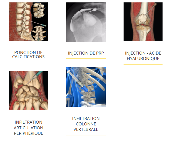
What are interventional radiology techniques?

Treating tendonitis with PRP
Pourquoi choisir IMPC pour votre prise en charge ?
IMPC propose une expertise spécialisée en radiologie interventionnelle ostéo-articulaire, dédiée au diagnostic et aux traitements mini-invasifs des pathologies osseuses et articulaires.
-
10 centres d’imagerie à Paris, facilement accessibles
-
Équipements de dernière génération, adaptés aux gestes interventionnels guidés par l’imagerie
-
Radiologues spécialisés en radiologie interventionnelle ostéo-articulaire
-
Rendez-vous rapides, sur Doctolib ou par téléphone
-
Compte rendu clair et rapide, disponible en ligne pour assurer la continuité des soins
