Pelvic MRI: a precise diagnosis of the pelvis

RDV IRM en ligne sur Doctolib
Prenez rendez-vous pour un IRM dans nos centres
Prise de rendez-vous IRM – Paris 75002
Prise de rendez-vous IRM – Paris 75009
Prise de rendez-vous IRM – Paris 75015
Prise de rendez-vous IRM – Paris 75009 Drouot

Comment se passe une IRM pelvienne dans notre centre ?
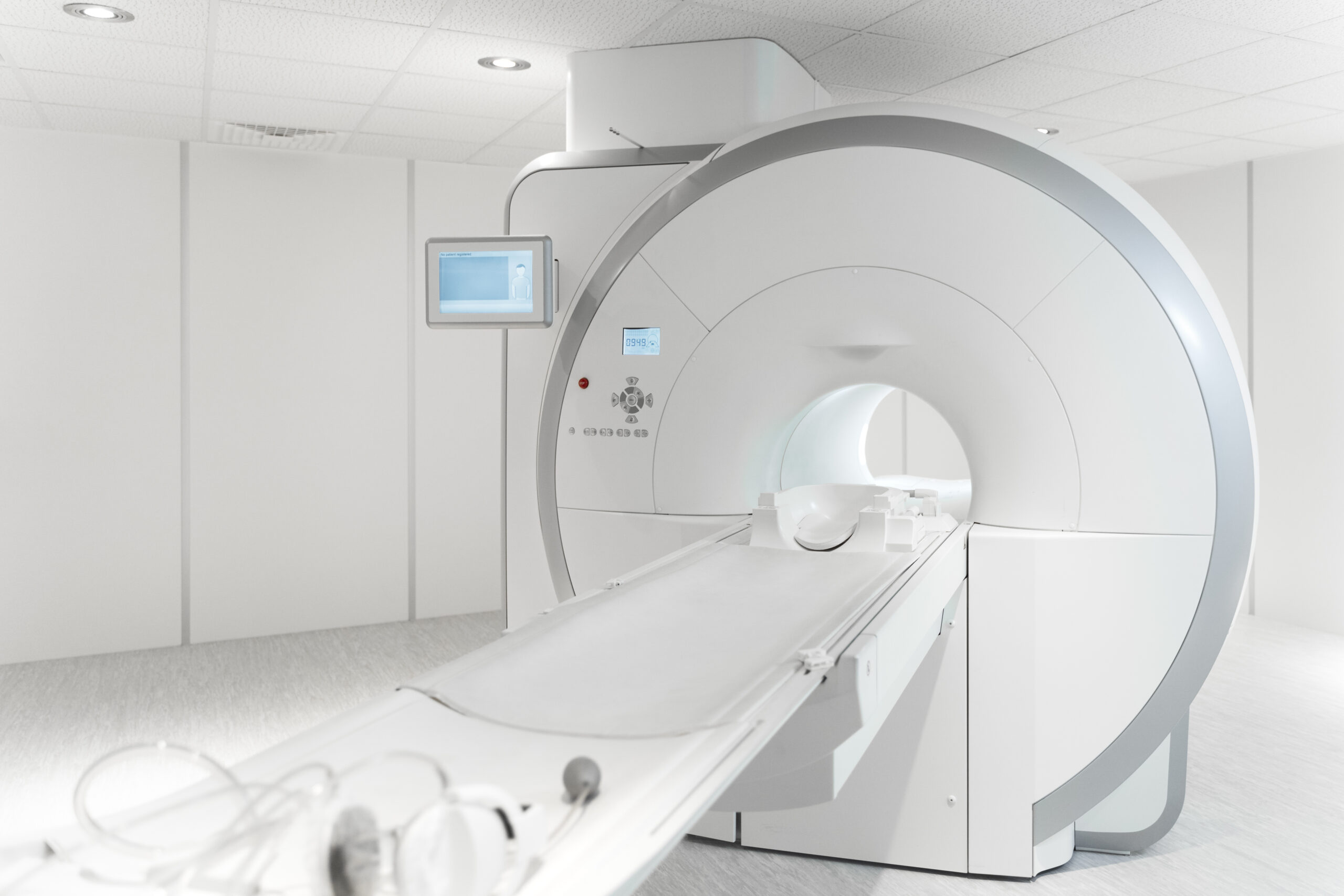
Cet examen se déroule dans un tunnel, la patiente est allongée sur le dos. La particularité de notre centre est que ce tunnel est semi-ouvert et que cette ouverture large permettra à nos patientes claustrophobes de passer aisément l’examen. Notre médecinspécialiste vous guidera durant toute la durée de l’examen.
Quelles sont les étapes de l’examen ?
- Le technicien manipulateur-radio vous appelle en salle d’attente, pour vous installer dans la cabine de préparation.
- Il vous est demandé d’enlever tout objet métallique sur vous (y compris piercing). Le soutien-gorge devra être retiré et vous resterez en culotte et tee-shirt pendant l’examen.
- Puis vous serez conduite dans la salle où se trouve la machine IRM afin de vous allonger sur la table d’examen.
- Une fois allongée, on vous mettra une antenne, c’est-à-dire une plaque afin de recueillir le signal.
- Après vous être rhabillée, vous retournerez en salle d’attente. Le médecin radiologue vous appellera dans les 20 mn pour vous donner une 1re interprétation des résultats DE L’IRM pelvienne.
- Vous partirez avec les clichés (papier et CD). Le compte-rendu vous est soit remis, soit téléchargeable sur notre site.
Particularité IMPC
Nos centres sont équipés d’IRM semi-ouvertes, facilitant l’examen pour les personnes claustrophobes.
L’IRM pelvienne est-elle efficace ?
Oui, l’IRM pelvienne est considérée comme un examen morphologique de référence.
- Pour l’endométriose, elle est le seul examen d’imagerie permettant un bilan complet.
- Elle fournit une cartographie précise des organes et de leurs éventuelles lésions.
- Elle dépasse largement l’échographie pour certaines indications, notamment les lésions profondes ou complexes.
Selon la Haute Autorité de Santé (2021), la précision diagnostique de l’IRM pelvienne dépasse 90 % pour certaines pathologies.
Douleurs pelviennes : quand consulter ?
Une douleur pelvienne est une douleur ressentie dans le bas-ventre, au niveau de l’utérus, de la vessie ou du rectum. Elle peut être sourde, intermittente ou aiguë.
Elle devient chronique lorsqu’elle est présente depuis plus de six mois, qu’elle soit cyclique ou non. Dans ce cas, une consultation médicale s’impose. Souvent, le médecin prescrit une imagerie comme l’IRM pelvienne pour en identifier la cause.
Endométriose : l’apport de l’IRM pelvienne
L’endométriose correspond à la présence de tissu endométrial (muqueuse utérine) en dehors de l’utérus. Trois formes existent :
- Endométriose ovarienne avec kystes (endométriomes).
- Endométriose péritonéale avec lésions multiples.
- Endométriose profonde atteignant les cloisons génitales ou le rectum.
Symptômes fréquents
- Règles douloureuses (dysménorrhées).
- Douleurs pendant les rapports sexuels.
- Troubles digestifs (diarrhées, constipation, ballonnements).
- Troubles de la fertilité.
Grâce à sa résolution élevée, l’IRM pelvienne est l’examen de choix pour localiser et mesurer les lésions d’endométriose, orientant ainsi la prise en charge médicale ou chirurgicale.
Étendue des explorations pelviennes par IRM
En gynécologie, l’IRM pelvienne visualise avec précision :
- les ovaires, la cavité utérine, le vagin,
- les voies urinaires et la vessie,
- le rectum et les organes digestifs voisins.
Elle est également utile en urologie (prostate, vessie) et en digestif (cancers colorectaux, malformations).
Elle peut guider une biopsie, une ponction ou une chirurgie.
Qu’est-ce qu’une IRM pelvienne ?
L’IRM pelvienne est un examen d’imagerie médicale non invasif qui explore avec précision les organes du bassin : utérus, ovaires, prostate, vessie, rectum. Contrairement au scanner, elle n’utilise pas de rayons X mais un champ magnétique et des ondes radio, ce qui la rend particulièrement sûre.
Il s’agit généralement d’un examen de deuxième intention, souvent prescrit après une échographie pelvienne pour affiner ou confirmer un diagnostic.
Grâce à son excellent contraste tissulaire, l’IRM pelvienne permet de détecter des pathologies complexes et d’apporter une aide essentielle au suivi thérapeutique.
Pourquoi faire une IRM pelvienne ?
APelvic MRI est un examen de référence dans de nombreuses situations médicales. Elle permet notamment de diagnostiquer :
- Endometriosis : touche environ 10 % des femmes en âge de procréer (Inserm, 2022). L’IRM est l’examen le plus précis pour établir un bilan complet et cartographier les lésions.
- Douleurs pelviennes chroniques : explorées lorsque les symptômes persistent depuis plus de six mois.
- Malformations utérines : utiles dans le bilan de fertilité.
- Pathologies ovariennes : kystes, polypes, syndrome des ovaires polykystiques (SOPK). L’IRM permet de classer les kystes dans des catégories de risque de cancer.
- Fibromes utérins et adénomyose : exploration précise pour adapter la prise en charge.
- Pathologies rectales ou vésicales : anomalies de la vessie ou du rectum, bilan d’extension de tumeurs digestives.
- Cancer de la prostate : en urologie, l’IRM pelvienne est incontournable pour le diagnostic et le suivi.
Une variante appelée IRM dynamique ou déféco-IRM peut être prescrite après une chirurgie ou en cas de troubles de la statique pelvienne. Elle évalue la mobilité des organes du petit bassin.
Préparation à l’examen
Pour optimiser la qualité des images, certaines consignes peuvent être données :
- Être à jeun pendant 4 à 6 heures dans certains cas.
- Ne pas vider complètement la vessie juste avant l’examen.
- Réaliser un lavement évacuateur 1 à 3 heures avant l’examen, notamment en cas de suspicion d’endométriose ou de pathologie rectale, afin d’éviter les artefacts d’image.
Injection de produit de contraste
Dans certains cas, une injection intraveineuse de gadolinium est réalisée. Elle permet de mieux visualiser certaines lésions ou tumeurs.
Les réactions allergiques sont extrêmement rares (<0,1 % des cas, HAS 2021).
Sécurité et contre-indications
L’IRM pelvienne est un examen sûr :
- aucun rayonnement ionisant,
- effets secondaires très rares,
- compatible avec la grossesse après le 1er trimestre (aucun effet nocif connu).
Contre-indications absolues
- Pacemaker non compatible.
- Implants auditifs ou certains dispositifs métalliques.
Tableau comparatif : IRM pelvienne vs échographie
| Critère | Pelvic MRI | Pelvic ultrasound |
|---|---|---|
| Précision diagnostique | Très élevée (>90 % pour endométriose) | Haute, dépend de l’opérateur |
| Rayons X | Aucun | Aucun |
| Duration | 20–40 minutes | 10–15 minutes |
| Indications principales | Endométriose, fibromes, cancers | Suivi simple, anomalies courantes, obstétrique |
Pourquoi choisir IMPC pour votre IRM pelvienne ?
En choisissant IMPC, vous bénéficiez de :
- a réseau de 10 centres d’imagerie médicale à Paris,
- from IRM de dernière génération,
- from radiologues spécialisés en gynécologie, urologie et digestif,
- a prise de rendez-vous rapide, en ligne ou par téléphone,
- un accompagnement humain et rassurant.
FAQ : Les questions les plus courantes sur l’IRM pelvienne
Peut-on faire une IRM pelvienne avec ses règles ?
Oui, c’est possible. Les règles ne faussent pas l’examen, mais selon le motif (ex. endométriose), le radiologue peut conseiller un moment précis du cycle.
Combien de temps dure une IRM pelvienne ?
L’examen dure en moyenne entre 20 et 40 minutes, selon la pathologie étudiée et l’éventuelle injection de produit de contraste.
Faut-il être à jeun avant une IRM pelvienne ?
Parfois. Un jeûne de 4 à 6 heures avant l’examen peut être demandé pour améliorer la qualité des images, surtout si un produit de contraste est utilisé.
L’IRM pelvienne est-elle sûre pour les femmes enceintes ?
Oui, après le premier trimestre. L’IRM n’utilise pas de rayons X et est considérée comme sûre pour les femmes enceintes.
Quand reçoit-on les résultats de l’IRM pelvienne ?
Le compte rendu et les images sont généralement disponibles le jour même. Ils peuvent aussi être consultés en ligne.
Quelles pathologies l’IRM pelvienne permet-elle de détecter ?
Elle explore notamment l’endométriose, les fibromes, les kystes ovariens, l’adénomyose, les cancers gynécologiques, les troubles de la statique pelvienne, ainsi que certaines pathologies de la vessie, du rectum et de la prostate.
Sources scientifiques
-
Inserm – Endométriose : mieux comprendre la maladie (2022)
-
Haute Autorité de Santé (HAS) – Imagerie par résonance magnétique, sécurité et indications (2021)
-
Société Française de Radiologie – Recommandations en imagerie pelvienne
Dernière mise à jour : le 30 août 2025
Dr Anne Elodie Millischer
