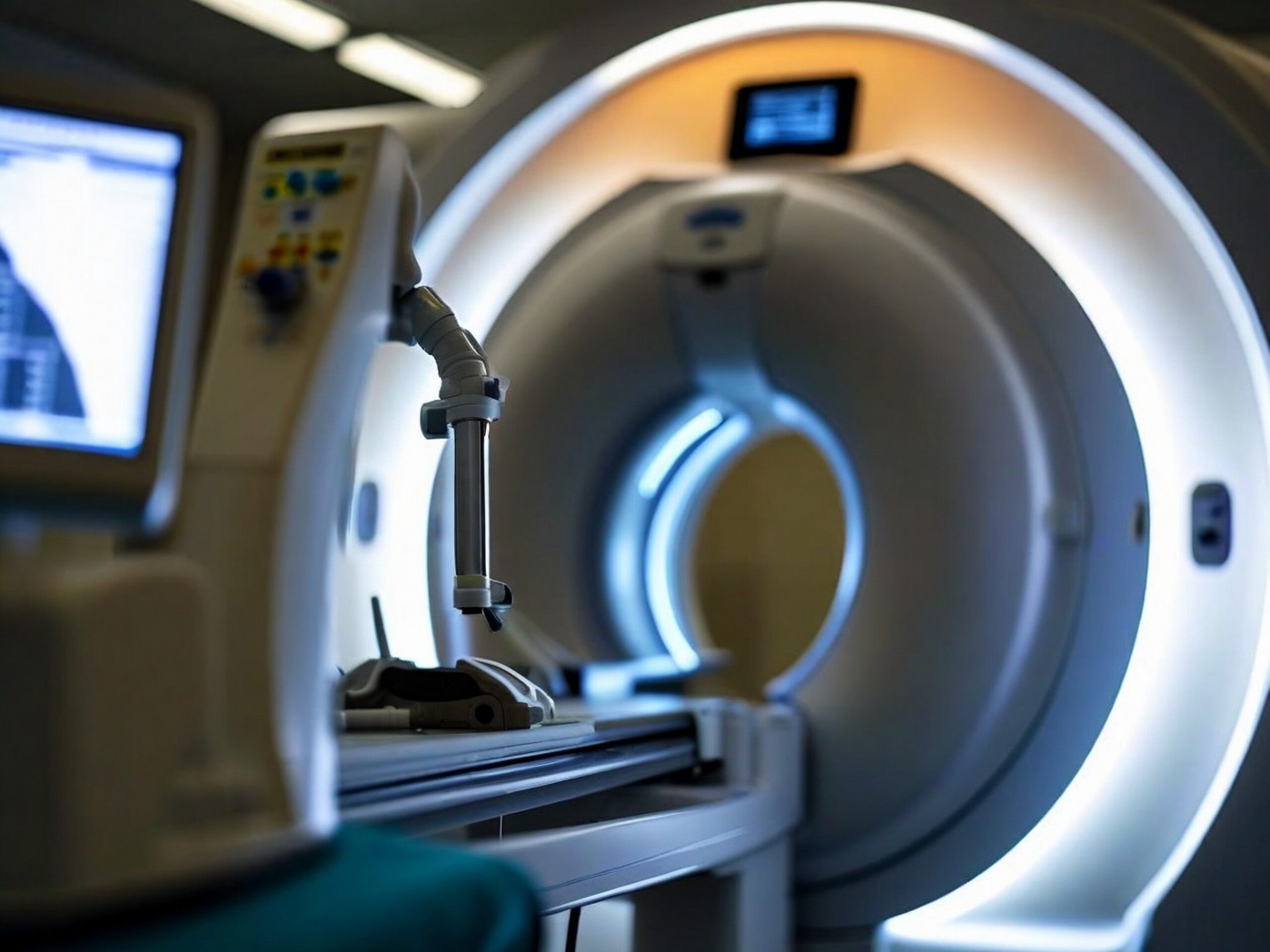
Chest Abdomen and Pelvis CT Scan

RDV scanner en ligne sur Doctolib
Prenez rendez-vous pour un scanner dans nos centres
Prise de rendez-vous SCANNER – Paris 75002
Prise de rendez-vous SCANNER – Paris 75009
Prise de rendez-vous SCANNER – Paris 75015

How does a TAP scan work?
Un scanner thoraco-abdomino-pelvien (TAP) est un examen d’imagerie très complet permettant d’explorer plusieurs régions clés du corps, comme le thorax, l’abdomen et le bassin. La plupart de ces scanners sont effectués avec l’administration d’un produit de contraste iodé injecté directement dans une veine. Ce produit améliore significativement la visibilité des structures internes, notamment les vaisseaux sanguins et les organes, et permet de mieux repérer d’éventuelles anomalies.
During the examination, the patient is positioned on a table that moves slowly through the scanner. It is important to remain still in order to obtain optimum image quality. In some cases, instructions such as holding your breath for a few seconds may be given.
The radiologist, a medical imaging specialist, chooses the most appropriate technique for your case. For example, depending on the nature of your symptoms or medical history, he or she may decide to perform thin sections or extend the exploration to other areas of the body. If necessary, this examination can be supplemented by other imaging techniques, such as ultrasound or MRI.
What is a virtual colonoscopy?
Virtual colonoscopy, also known as colonoscanner, is an advanced diagnostic method using X-rays to examine the colon. Unlike traditional colonoscopy, it does not require the insertion of an endoscope into the colon, making it less invasive and more comfortable for the patient.
As with a conventional CT scanner, the device collects detailed images of the inside of the abdomen, taken from different angles. These images are then processed by sophisticated computer software, which assembles them to produce a highly accurate 3D model of the inside of the colon.
Before the examination, a colonic preparation is often required to clean the colon and ensure clear images. During the examination, a small amount of air or CO₂ may be insufflated to better visualize the colon walls.
Why perform a virtual colonoscopy?
This technique is particularly useful for detecting precancerous lesions, such as polyps, in asymptomatic patients or those at increased risk of colorectal cancer. It is often proposed when conventional colonoscopy is contraindicated, for example due to health problems or complex anatomy.
It can also be performed when a conventional colonoscopy has not been completed, for example if a full colonic examination has been interrupted. It is also an interesting alternative for patients reluctant to undergo an invasive examination.
Virtual colonoscopy is frequently recommended after a positive fecal occult blood test, to determine the origin of the bleeding. Its effectiveness as a screening tool makes it a valuable method for identifying pathologies at an early stage.
What are the most frequent anomalies that can be detected by a CAP CT Scan ?
The scanner's usefulness is extensive, since it can be used to :
- Préciser un diagnostic d’une maladie localisée.
- Monitor the evolution of a disease under treatment.
- To perform a check-up before or after surgery. It usually requires an iodine injection to better study the organs and pathology.
In addition, it is systematically recommended for the initial assessment and follow-up of TG :
- Abdomen & pelvis : the CT scan is characterized by its sensitivity of 80 % for the analysis of retroperitoneal adenopathies.
- Chest : this is the most sensitive examination for detecting pulmonary metastases and mediastinal adenopathies.
As mentioned, this is a inescapable to diagnose certain cancers.
Can a CAP CT scan diagnose cancer?
The creation of a CAP CT Scan est depuis plusieurs années l’examen de référence utilisé pour rechercher les cancers occultes chez les patients.
The Chest Abdomen and Pelvis CT scan (CAP) is recommended in many cases and can detect, among other things, anomalies such as :
- Traumatic injury to the liver, spleen, kidneys
- Traumatic injury to the pancreas (par exemple, abcès, tumeur primaire ou secondaire).
- Traumatic injury to the pelvic organs
Ce type d’examen va permettre le diagnostic des cancers liés à l’utérus ou à la prostate
TAP scan with injection
This examination is often performed with an intravenous injection of an iodinated contrast medium, enabling precise visualization of internal organs, blood vessels and surrounding structures. Thanks to this technique, pathologies such as tumors, inflammation, infections, vascular anomalies or traumatic lesions can be detected. The patient lies on a table that moves through the scanner, and breathing instructions can be given to ensure sharp, precise images. This fast, non-invasive examination is an essential tool for establishing a reliable diagnosis and guiding appropriate treatment.
What is a Chest Abdomen and Pelvis CT scan?
A Chest Abdomen and Pelvis CT Scan is to explore the entire chest, abdomen and pelvis area. This examination is used in the diagnosis and follow-up of tumoral, infectious or inflammatory pathologies.
That said, it plays a leading role in the cancer diagnosis and follow-up. It is decisive in confirming or disqualifying a pathology.
Scanner thoraco-abdomino-pelvien : pourquoi ?
Pour découvrir ou exclure une pathologie grave, identifier l’étiologie de ces plaintes douloureuses, prendre une décision thérapeutique ou mieux définir un pronostic et, finalement, répondre à une anxiété du patient ou personnelle. Par ailleurs, cet examen a le mérite d’examiner plusieurs organes.
What organs are involved in a CAP scan?
The Chest Abdomen and Pelvis CT scan can be used to visualize and examine the lungs, mediastinum (région de la cage thoracique située entre les deux poumons contenant le cœur, l’œsophage, la trachée et les deux bronches souches). De surcroît, il accorde une attention particulière aux gros vaisseaux sanguins et lymphatiques ainsi qu’aux nerfs qui y passent également, au tube digestif (du bas œsophage au rectum), aux organes pleins (foie, rate, pancréas, reins), aux vaisseaux et aux ganglions de l’abdomen et du pelvis.
In this sense, visualizing these organs can reveal certain anomalies.
Preparation for a TAP scanner
Some scanners require special preparation beforehand.
For most examinations, you will need to drink and/or receive an injection of contrast material. This is a dye that helps to visualize body tissues more clearly on the scanner. The injection is made through a small, thin tube (cannula) in your arm. The cannula remains in place until the end of your examination, in case you experience problems such as an allergic reaction after the injection.
CT scans of the abdomen
If you have a CT scan of your abdomen, you may need to :
- boire un produit de contraste liquide un certain temps avant le scanner ;
- boire davantage de ce produit ou de l’eau dans le service de radiologie.
- arrêter de manger ou de boire après minuit la nuit précédant le scanner (cela concerne un scanner de l’intérieur du gros intestin, appelé colographie CT).
You will usually receive the contrast medium by injection and also as a drink. This helps to better visualize the digestive system on the scanner.
Chest scans
You may receive an injection of contrast material during the scan. This helps to better visualize tissues close to the area containing the cancer. For example, if your doctor wants to know if the cancer is affecting your blood vessels. This can help determine whether or not the cancer can be surgically removed.
Pelvic scanners
The pelvis is the lower body cavity located between the hip bones. It contains the pelvic organs, including the bladder, lower large intestine and reproductive organs. If you have a pelvic CT scan, you may need to :
- arrêter de manger ou de boire pendant un certain temps avant le scanner.
- recevoir une injection de produit de contraste.
Sometimes, for a rectal examination, an enema with a contrast medium is necessary. This appears on the X-ray and helps to better visualize the outline of the intestine on the CT scan. This may cause constipation. Your first stools will be white, but there are no other side effects.
