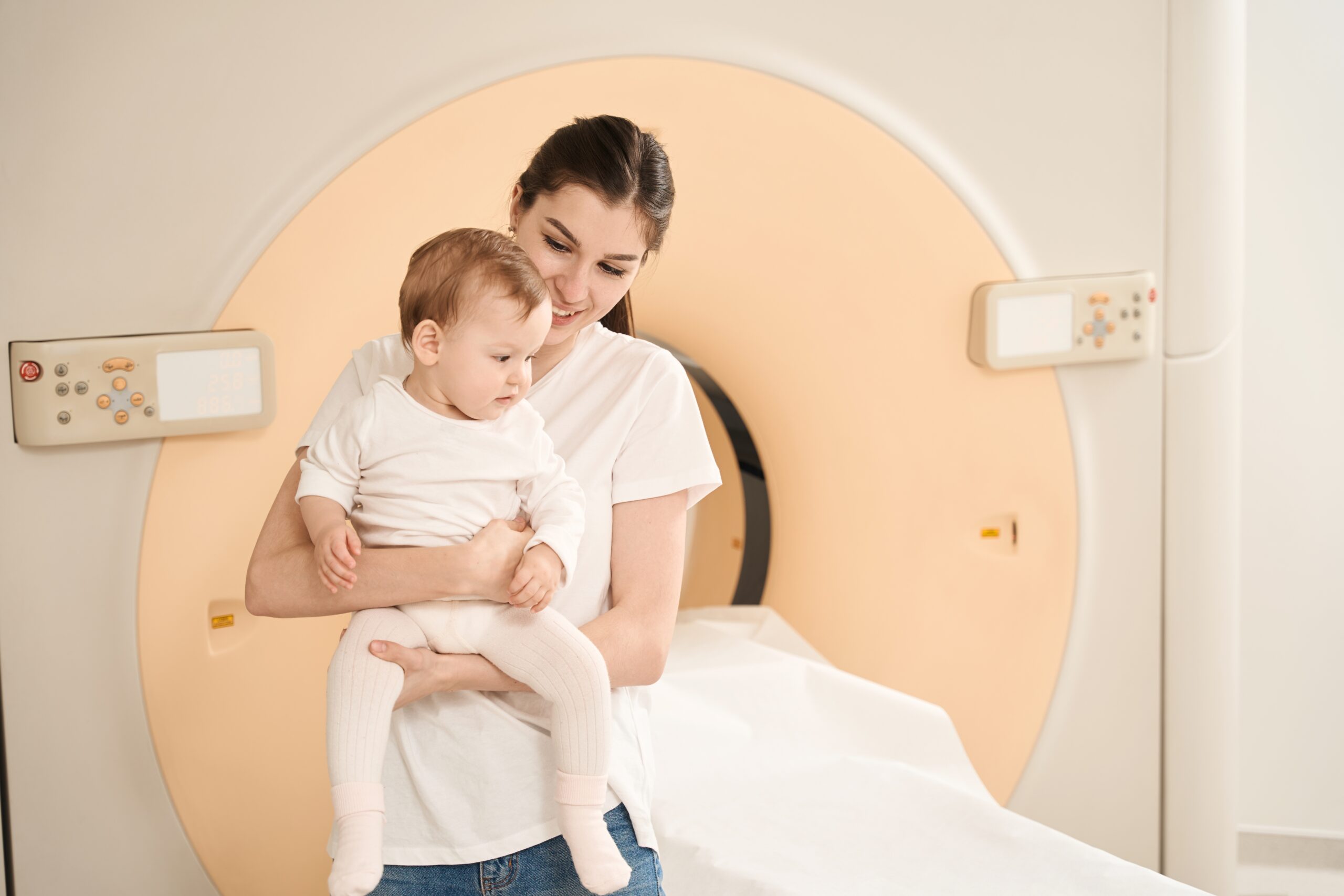
Imaging of women and children
Imaging women and children: an IMPC centre of excellence

RDV en ligne sur Doctolib
Prenez rendez-vous pour un échographie dans nos centres
Prise de rendez-vous – Paris 75009
Prise de rendez-vous – Paris 75015
Prise de rendez-vous – Paris 75009 Drouot

Imaging center for women and children
The concern for uniqueness has motivated the development of the Mother-Child Imaging Center. This center offers the skills of several specialist radiologists, who can act as experts:
- in gynecological imaging from adolescence through to post-menopause: for expertise on cysts, bleeding, menstrual disorders, endometriosis, fertility exploration, menopausal follow-up, whether in ultrasoundhysterography and/or MRI ;
- on the senological level, both for mammographic screening and assessment of breast masses and pain, whether by mammography, tomosynthesis or breast MRI;
- obstetrics: pregnancy follow-up, referral advice, with all radiologists practicing in this field, being an integral part of Parisian hospital-based prenatal diagnosis centers (Necker - Port Royal - Robert Debré) and/or affiliated to an obstetric imaging network
- in paediatrics: both for the monitoring of newborns, concerning pediatric radiographs lung, pelvis, abdomen without preparation, the echograms hip, medullary, renal and trans-frontanellar ultrasound.
Each patient will have the possibility of having all her examinations grouped together in a single computer file, even if they are carried out in different geographical locations of our center, archived over several decades.
A qualified team for pediatric radiology
Our pediatric radiology is designed with children in mind, whether it's our child-specific technology pediatrics or our child-friendly technical platform. Our state-of-the-art equipment enhances image quality while using the latest low-dose radiation technology. Our radiologists are specially trained in pediatrics, providing expert interpretation and diagnosis.
Pediatric imaging: state-of-the-art technology for a reliable examination
In addition, we are equipped to add the BLADE sequence to our MRI protocols. This new MR acceleration and motion correction technique enables us to perform free-breathing abdominal MRI in children, reduce motion artifacts in MR scans, and reduce MR scan times and sedation requirements.
The women's imaging center
Our women's imaging center provides personalized follow-up and diagnosis for each of our patients. This is achieved through a wide range of examinations, some of which are listed below.
Obstetrical ultrasound
L'obstetrical ultrasound uses sound waves to produce baby pictures (embryo or fetus) within the pregnant woman, as well as the mother's uterus and ovaries. It does not use ionizing radiation, has no known harmful effects and is the preferred method for monitoring pregnant women and their unborn baby.
L'obstetrical ultrasound is a useful clinical examination for :
- establish the presence of a living embryo/foetus
- estimate age of pregnancy
- diagnose congenital anomalies of the fetus
- assess fetal position
- assess the position of the placenta
- identify multiple pregnancies
- determine the amount of amniotic fluid around the baby
- check opening or shortening of the cervix
- assess fetal growth
- assess fetal well-being
Pelvic MRI
L'Pelvic MRI creates images of the area between your hips. It can detect problems with the pelvic bones, intestines, bladder, reproductive organs, lymph nodes, etc.
Abnormal results following Pelvic MRI for women may suggest :
A cervical cancerof the endometrium or ovaries.
- An adenomyosis, when tissues similar to the uterine lining attach to the muscular wall of the uterus.
- Congenital anomalies of the reproductive organs
- The world of medical imaging has made significant strides in diagnosing and understanding the human body. As one of these technologies, Magnetic Resonance Imaging (MRI) offers detailed images of internal structures without requiring invasive procedures. In this article, we discuss the crucial role of lumbar spine MRI in diagnosing conditions affecting the lower back. endometriosiswhen tissue similar to the uterine mucosa develops outside the uterus in areas such as the Fallopian tubes and ovaries.
- From ovarian growths
- Structural problems of the reproductive organs
- Uterine fibroids are non-cancerous growths that appear on your uterus.
Pelvic ultrasound
The world of medical imaging has made significant strides in diagnosing and understanding the human body. As one of these technologies, Magnetic Resonance Imaging (MRI) offers detailed images of internal structures without requiring invasive procedures. In this article, we discuss the crucial role of lumbar spine MRI in diagnosing conditions affecting the lower back. pelvic ultrasound is an examination that uses sound waves to obtain images of the organs in your pelvis. Your doctor may order this test to diagnose an illness or to check on your baby's health while still in the womb.
For women, thepelvic ultrasound is used to view the following items:
- the cervix
- visit Fallopian tubes
- ovaries
- the uterus
- the Vagina
- or the Bladder
Women's centers

MOTHER-CHILD IMAGING CENTER
The woman-mother-child imaging center was built around the idea of a continuum between these different stages that a woman may encounter. Our center brings together different radiologist-specialists capable of intervening specifically at each of these stages: a radiologist specialized in gynecological and senological imaging of women, a radiologist specialized in obstetrical ultrasound, and a radiopediatrician specialized in pediatric imaging from the neonatal period through to adolescence.
For more information, visit the dedicated website:

UTERINE FIBROIDS CANCEROLOGY UNIT
The fibroma center was built with a dual objective in mind:
- Inform patients and professionals about the most modern, least invasive and most appropriate medical and therapeutic solutions for every situation.
- Treat uterine fibroids and other pathologies by embolization, with a team specialized in the field. Our interventional radiologists have been practicing embolization for years now.
For more information, visit the dedicated website:
